Medical Image Processing
m |
m (→Links) |
||
| Line 30: | Line 30: | ||
* [http://www.shu.ac.uk/mmvl/research/research-active-Medical.html Official MMVL Medical Image Processing page] | * [http://www.shu.ac.uk/mmvl/research/research-active-Medical.html Official MMVL Medical Image Processing page] | ||
* [http://vtk.org/ The Visualisation Toolkit] | * [http://vtk.org/ The Visualisation Toolkit] | ||
| − | |||
[[Category:Projects]] | [[Category:Projects]] | ||
[[Category:Medical Image Processing|*Medical Image Processing]] | [[Category:Medical Image Processing|*Medical Image Processing]] | ||
Revision as of 14:25, 12 March 2009
Contents |
Medical Image Processing
3D data processing
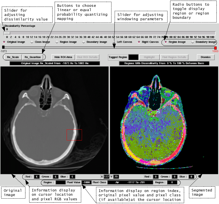
The MMVL is doing research on 3D Medical Image Processing. Exspecially for processing of 3D data, the Visualisation Toolkit has proven to be useful. Iso Surface Extraction is an example, which demonstrates, how powerful VTK is.
Ultrasound
Equipment:
Portable PC Based Ultrasound machine: Echo Blaster 128 INT-2Z Kit
Convex Ultrasound probe(3MHz to 7MHz): C4.5/50/128Z
On going projects
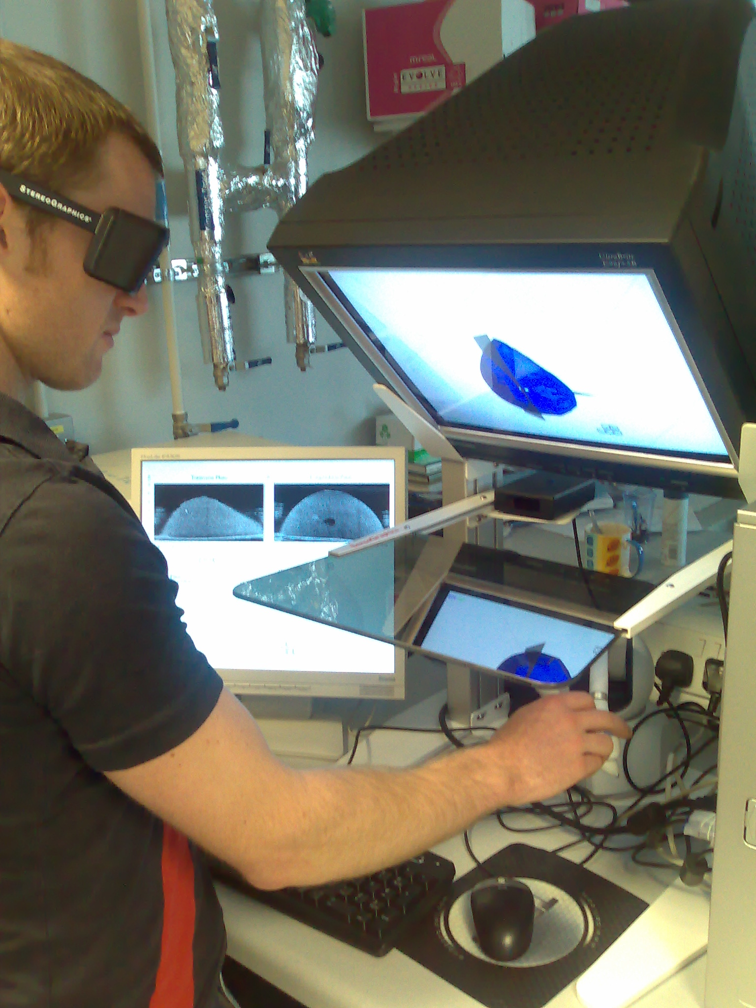
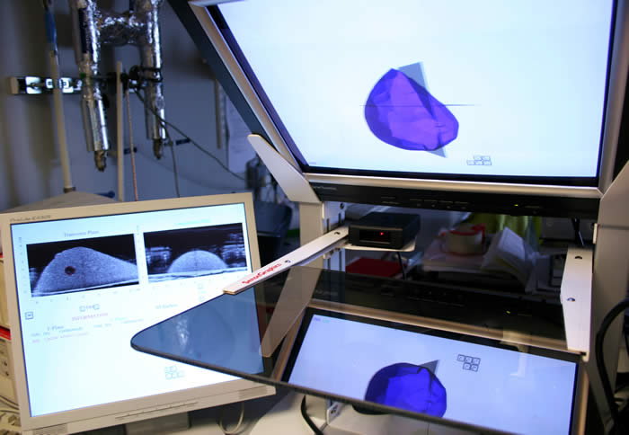
Medical Ultrasound Training Simulation Using Haptic Force Feedback Mechanism
(By MSc student, Jin Quan Tissa Tan, Supervisor: Arul N. Selvan )
The aim of the project was to create a highly realistic (haptic-based) medical ultrasound training simulator for training a sonographer. The purpose of this simulator is to provide the sense of touch to the user and reconstruct the anatomy as a model using the actual ultrasound data.


