Microscope Environment
m |
m |
||
| (3 intermediate revisions by one user not shown) | |||
| Line 1: | Line 1: | ||
| + | <html> | ||
| + | <div class="thumb tright"><div style="width:400px;"> | ||
| + | <object type="application/x-shockwave-flash" data="http://vision.eng.shu.ac.uk/jan/flv/flvplayer.swf" width="400" height="364"> | ||
| + | <param name="flashvars" | ||
| + | value="file=http://vision.eng.shu.ac.uk/jan/flv/sugar.xml&searchbar=false&displayheight=300" /> | ||
| + | <param name="movie" value="http://vision.eng.shu.ac.uk/jan/flv/flvplayer.swf" /> | ||
| + | <param name="allowfullscreen" value="true" /> | ||
| + | </object> | ||
| + | <div class="thumbcaption">Demo of automated positioning of micro-object (here: a piece of sugar)</div> | ||
| + | </div></div> | ||
| + | </html> | ||
=MiCRoN Tests With Microscope= | =MiCRoN Tests With Microscope= | ||
==Recognition/Tracking the Syringe Chip== | ==Recognition/Tracking the Syringe Chip== | ||
| Line 11: | Line 22: | ||
{|align=center | {|align=center | ||
|- | |- | ||
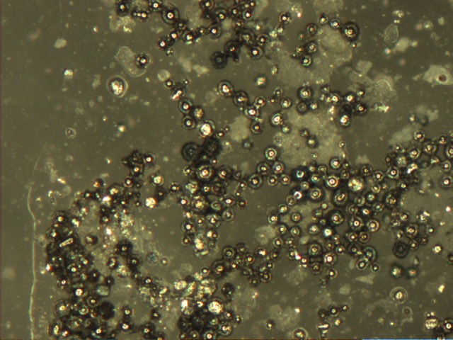
| − | + | |[[Image:Solder.jpg|thumb|250px|Microscope image of small solder spheres]]|||[[Image:Sugarsetup.jpg|thumb|250px|Setup for automatically aligning a piece of sugar with the camera coordinate system (6.3 MByte [http://vision.eng.shu.ac.uk/jan/automated.avi video])]]||[[Image:Sugarpush.jpg|thumb|250px|Automated tungsten tip approaching the piece of sugar (6.7 MByte [http://vision.eng.shu.ac.uk/jan/sugarpush.avi video])]] | |
|- | |- | ||
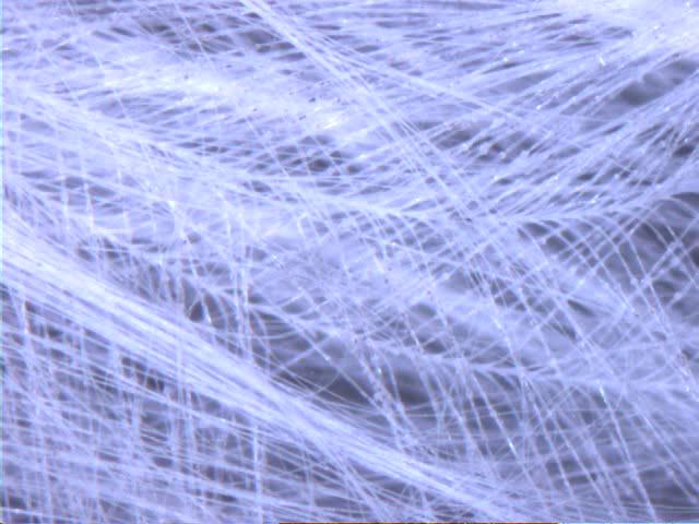
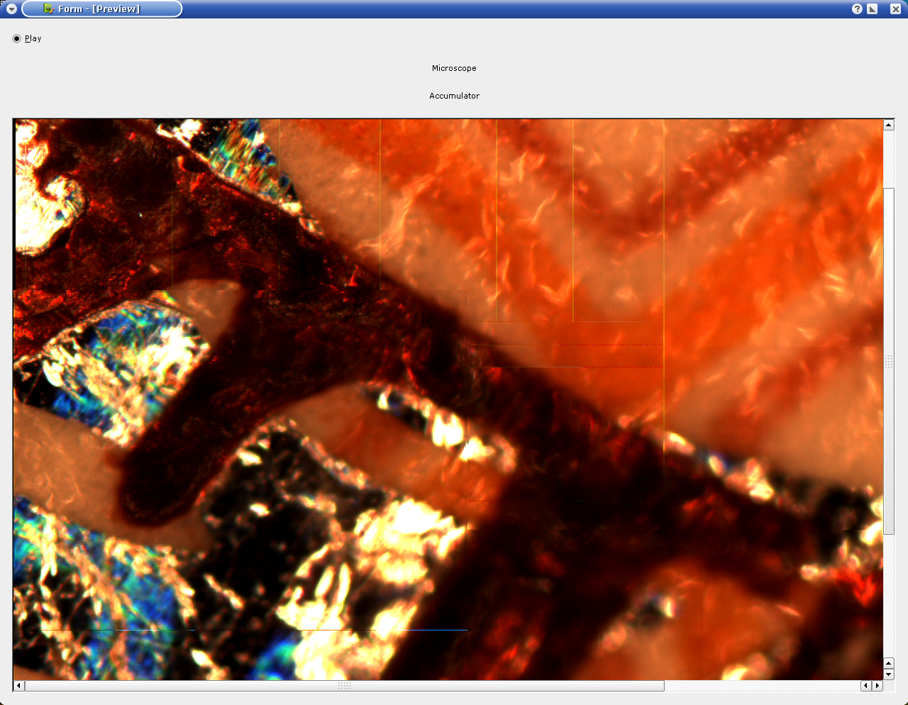
|[[Image:Feather.jpg|thumb|250px|A bird's feather (reflected light, darkfield) (7.2 MByte [http://vision.eng.shu.ac.uk/jan/feather1.avi video], 10.1 MByte [http://vision.eng.shu.ac.uk/jan/feather2.avi video])]]||[[Image:Milk.jpg|250px|thumb|Microscope image of milk (9.5 MByte [http://vision.eng.shu.ac.uk/jan/milk.avi video])]]||[[Image:Mapping.png|250px|thumb|Lateral stitching using the feedback of the microscope's drive]]|| | |[[Image:Feather.jpg|thumb|250px|A bird's feather (reflected light, darkfield) (7.2 MByte [http://vision.eng.shu.ac.uk/jan/feather1.avi video], 10.1 MByte [http://vision.eng.shu.ac.uk/jan/feather2.avi video])]]||[[Image:Milk.jpg|250px|thumb|Microscope image of milk (9.5 MByte [http://vision.eng.shu.ac.uk/jan/milk.avi video])]]||[[Image:Mapping.png|250px|thumb|Lateral stitching using the feedback of the microscope's drive]]|| | ||
| Line 18: | Line 29: | ||
=See Also= | =See Also= | ||
| + | * [[Microscope Vision Software]] | ||
* [[Microscope Control Software]] | * [[Microscope Control Software]] | ||
* [[Micron|MiCRoN project]] | * [[Micron|MiCRoN project]] | ||
| − | |||
* [[Pipette Reconstruction]] | * [[Pipette Reconstruction]] | ||
| + | [[Category:Projects]] | ||
[[Category:Micron]] | [[Category:Micron]] | ||
Latest revision as of 18:05, 6 February 2011
Contents |
[edit] MiCRoN Tests With Microscope
[edit] Recognition/Tracking the Syringe Chip
The same software, which was applied in the MiCRoN test environment also can be applied to a microscope. The video input in this case was a firewire digital camera (IIDC/DCAM compatible). At the moment we are using a motorized Leica DM LAM controlled by serial-port and a Basler A302fc IIDC/DCAM compatible firewire camera.
[edit] Rotating A Piece Of Sugar
If you have a motorized microscope, a fast camera and a computer, you just need vision software to do robotics under your microscope. Semi-automated tools, which are assisting the human operator, are conceivable. Here you can see a proof-of-concept for this.
After the piece of sugar and the tungsten tip have been brought into field of view manually, the system automatically aligns the piece of sugar with the coordinate system of the camera. If you watch the video, you'll see, that every time the piece of sugar is lost by the vision system, the tungsten tip will be moved to a neutral position.
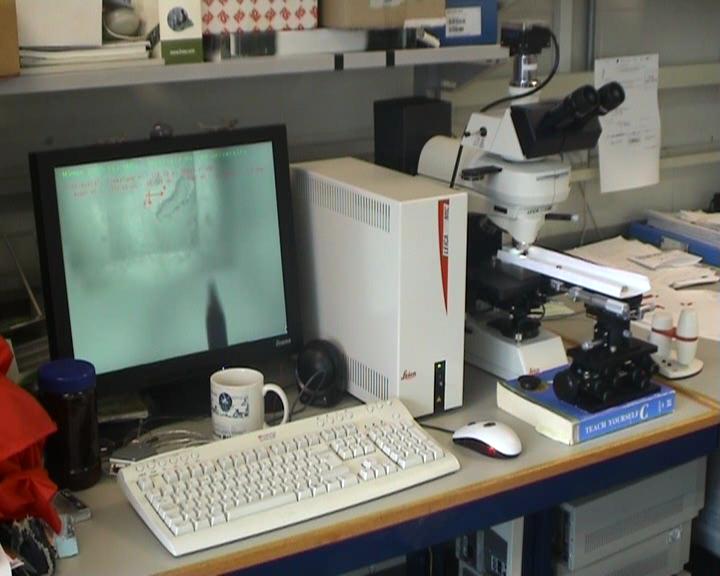 Setup for automatically aligning a piece of sugar with the camera coordinate system (6.3 MByte video) |
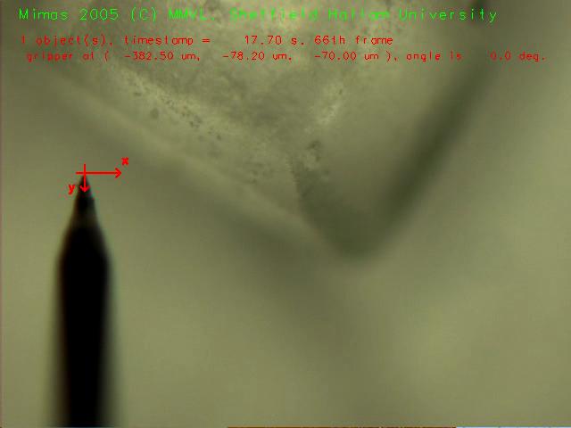 Automated tungsten tip approaching the piece of sugar (6.7 MByte video) | ||
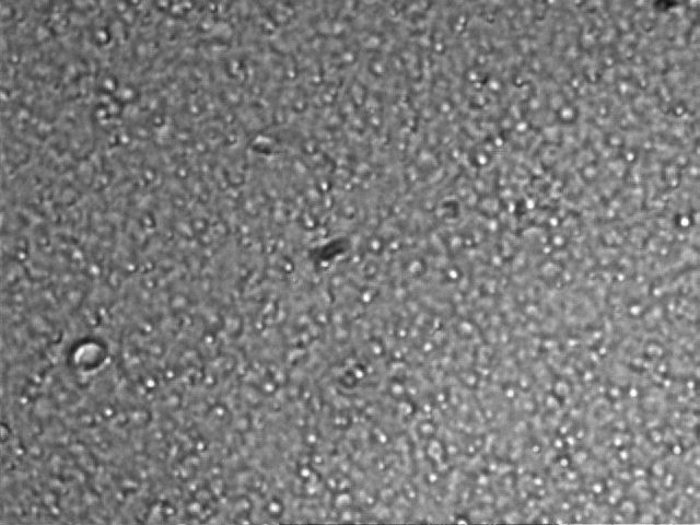 Microscope image of milk (9.5 MByte video) |
