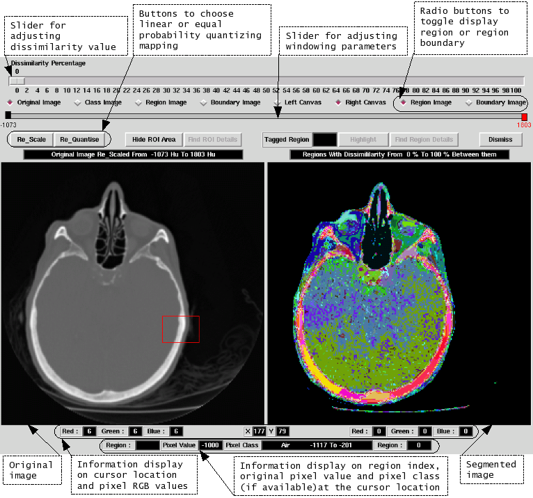
Medical Image Processing
From MMVLWiki
(Difference between revisions)
(→3D data processing) |
m (→Links) |
||
| (23 intermediate revisions by 3 users not shown) | |||
| Line 1: | Line 1: | ||
| − | {|align= | + | =Medical Image Processing= |
| + | |||
| + | {| | ||
| + | {|align="center" | ||
|- | |- | ||
| − | |[[Image:MedicalSegmentationGui.png|thumb| | + | |[[Image:MedicalSegmentationGui.png|thumb|221px|GUI for displaying hierarchical segmentation results to detect '''stroke lesions''']] |
| − | | | + | ||[[Image:Medical_Ultrasound_Training_Simulator1.jpg|thumb|155px|Medical ultrasound training simulator in operation]] |
| − | |[[Image: | + | ||[[Image:001general image.jpg|thumb|300px|Close up of the medical ultrasound training simulator system]] |
|- | |- | ||
|} | |} | ||
| − | + | =Ultrasound= | |
| − | + | ||
| − | + | ||
| − | + | ||
| − | + | ||
| − | + | ||
| − | + | ||
| − | + | ||
Equipment: | Equipment: | ||
| Line 20: | Line 16: | ||
Convex Ultrasound probe(3MHz to 7MHz): [http://www.telemed.lt/prb_scan_eb128_en.htm C4.5/50/128Z] | Convex Ultrasound probe(3MHz to 7MHz): [http://www.telemed.lt/prb_scan_eb128_en.htm C4.5/50/128Z] | ||
| + | |||
| + | =On going projects= | ||
| + | '''Medical Ultrasound Training Simulation Using Haptic Force Feedback Mechanism''' | ||
| + | |||
| + | (By MSc student, Jin Quan Tissa Tan, Supervisor: [mailto:A.N.Selvan@REMOVETHISshu.ac.uk Arul N. Selvan] ) | ||
| + | |||
| + | The aim of the project was to create a highly realistic (haptic-based) medical ultrasound training simulator for training a sonographer. The purpose of this simulator is to provide the sense of touch to the user and reconstruct the anatomy as a model using the actual ultrasound data. | ||
=Links= | =Links= | ||
* [http://www.shu.ac.uk/mmvl/research/research-active-Medical.html Official MMVL Medical Image Processing page] | * [http://www.shu.ac.uk/mmvl/research/research-active-Medical.html Official MMVL Medical Image Processing page] | ||
* [http://vtk.org/ The Visualisation Toolkit] | * [http://vtk.org/ The Visualisation Toolkit] | ||
| + | * [http://diamond.cs.cmu.edu/ OpenDiamond for assisting in diagnosis of breast cancer] | ||
[[Category:Projects]] | [[Category:Projects]] | ||
[[Category:Medical Image Processing|*Medical Image Processing]] | [[Category:Medical Image Processing|*Medical Image Processing]] | ||
Latest revision as of 09:31, 27 July 2009
Contents |
[edit] Medical Image Processing
[edit] Ultrasound
Equipment:
Portable PC Based Ultrasound machine: Echo Blaster 128 INT-2Z Kit
Convex Ultrasound probe(3MHz to 7MHz): C4.5/50/128Z
[edit] On going projects
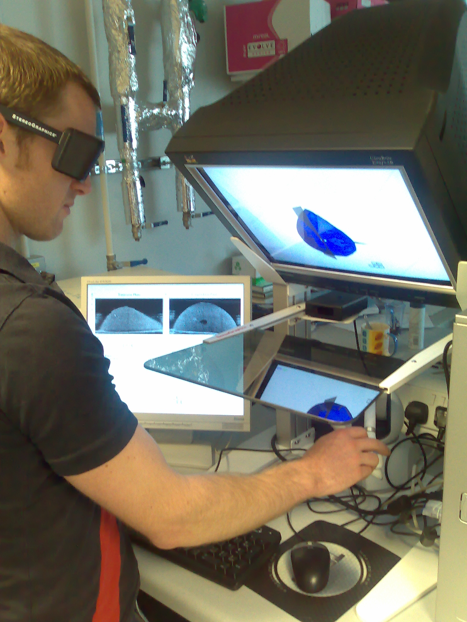
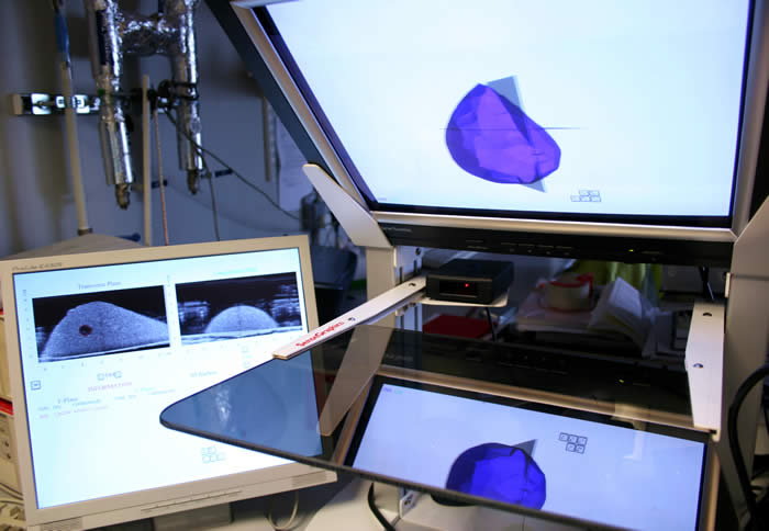
Medical Ultrasound Training Simulation Using Haptic Force Feedback Mechanism
(By MSc student, Jin Quan Tissa Tan, Supervisor: Arul N. Selvan )
The aim of the project was to create a highly realistic (haptic-based) medical ultrasound training simulator for training a sonographer. The purpose of this simulator is to provide the sense of touch to the user and reconstruct the anatomy as a model using the actual ultrasound data.
[edit] Links
- Official MMVL Medical Image Processing page
- The Visualisation Toolkit
- OpenDiamond for assisting in diagnosis of breast cancer


