Microscope Environment
m |
m |
||
| Line 18: | Line 18: | ||
* [[Microscope Control Software]] | * [[Microscope Control Software]] | ||
* [[Micron|MiCRoN project]] | * [[Micron|MiCRoN project]] | ||
| − | * [[ | + | * [[Microscope Vision Software]] |
* [[Pipette Reconstruction]] | * [[Pipette Reconstruction]] | ||
[[Category:Micron]] | [[Category:Micron]] | ||
Revision as of 13:45, 10 March 2006
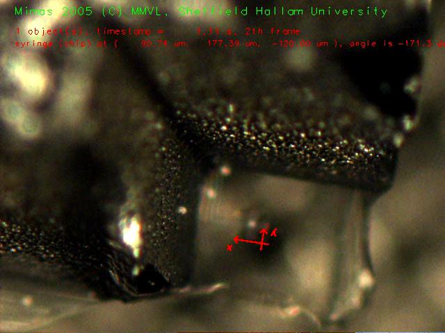
 Object recognition and tracking of syringe chip in 3-D/4 DOF (16 MByte video) |
|
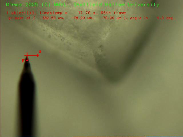
 Setup for automatically aligning a piece of sugar with the camera coordinate system (6.3 MByte video) |
 Automated tungsten tip approaching the piece of sugar (6.7 MByte video) |
Contents |
MiCRoN Tests With Microscope
Recognition/Tracking the Syringe Chip
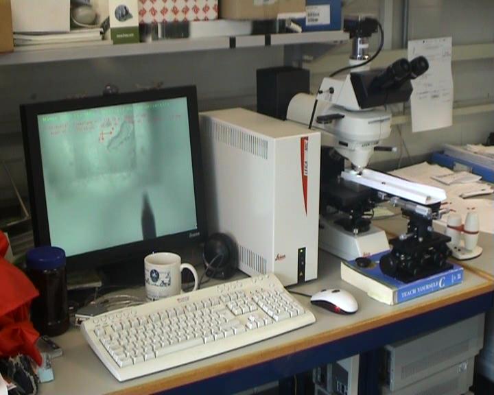
The same software, which was applied in the MiCRoN test environment also can be applied to a microscope. The video input in this case was a firewire digital camera (dc1394).
Rotating A Piece Of Sugar
If you have a motorized microscope, a fast camera and a computer, you just need vision software to do robotics under your microscope. Semi-automated tools, which are assisting the human operator, are conceivable. Here you can see a proof-of-concept for this.
After the piece of sugar and the tungsten tip have been brought into field of view manually, the system automatically aligns the piece of sugar with the coordinate system of the camera. If you watch the video, you'll see, that every time the piece of sugar is lost by the vision system, the tungsten tip will be moved to a neutral position.
