Microscope Control Software
From MMVLWiki
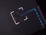
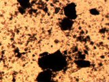
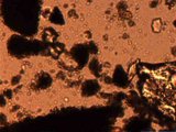
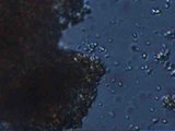
Microscope video (FOV width 500 um) of eucaryote (934kB Divx3-video)
| Table of contents |
[edit]
Microscope Control Software
[edit]
Hardware
At the MMVL a Linux software for controlling a Leica DM LAM microscope with a Basler A302fc IIDC/DCAM-compatible firewire camera has been developed.
[edit]
Software
[edit]
Features
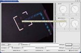
The software uses OpenGL to display the camera image with a high framerate. The software already has the following capabilities:
- Move object table with mouse-dragging
- Camera-display with digital zoom (requires IIDC 1394-based firewire digital camera)
- Display and capture videos:
- Colour images
- Graylevel images
- Edge images (thresholded Sobel)
- Capture focus stacks
[edit]
Implementation
The implementation took maybe 15 days. Before being able to develop the application itself, the Mimas-library had to be enhanced with firewire digital camera input, the existing libserial (http://sourceforge.net/projects/libserial/)-library had to be enhanced with timing functionality and the required part of the serial communication with the Leica DM LAM microscope had to be implemented under Linux.
[edit]
Download
- You need to have
 Qt3 (http://www.trolltech.com/) and libdc1394 (http://sourceforge.net/projects/libdc1394/) on your computer.
- You need to install
 libserial-0.4.1.tar.gz (http://vision.eng.shu.ac.uk/jan/libserial-0.4.1.tar.gz) (195 kByte).
- You also need
 Mimas-1.4 (http://vision.eng.shu.ac.uk/jan/mimas/mimas-1.4.tar.bz2) (12.2 MByte).
- Finally you need to install leica.tar.gz (http://vision.eng.shu.ac.uk/jan/leica.tar.gz) (32 kByte).
[edit]
Gallery
 |  |  |  |  |
[edit]
See Also

[edit]
External Links
- European Microscopy Site (http://www.micro-scope.de/toc.html)
- Leica Microsystems (http://www.leica-microsystems.com/) (on this site you can find the documentation of the serial protocol)
- David Attenborough - Life In The Undergrowth (http://www.bbc.co.uk/sn/tvradio/programmes/lifeintheundergrowth/)

